44 dissecting microscope diagram with labels
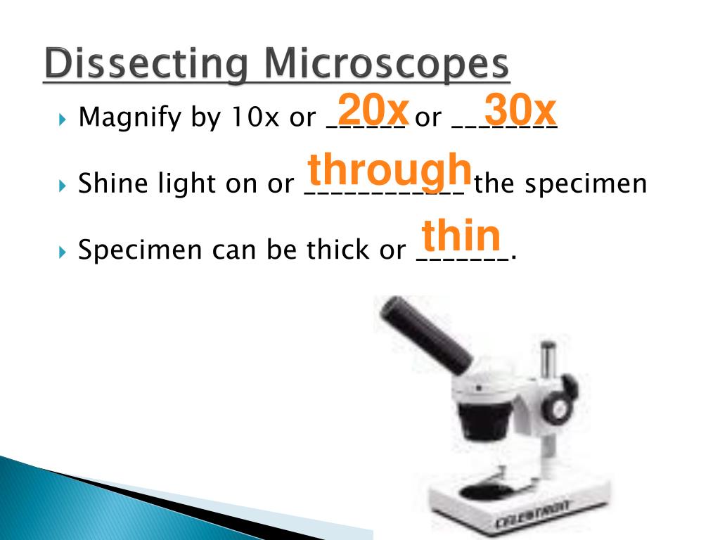
Parts of the Dissecting Microscope - Synonym Dissecting microscopes are used for viewing live specimens or three-dimensional objects too large or thick to be accommodated by compound microscopes. Specimens can be physically manipulated under magnification, since they do not have to be mounted onto a slide for observation under a dissecting microscope. These ... Dissecting microscope (Stereo or stereoscopic microscope)- Definition ... Stereo Zoom Dissecting Microscope-They are trinocular or binocular dissecting microscopes with a zooming range of 6.7x-45x. They can be attached to the digital camera which takes photos of the viewing images. They have a dual-LED illuminator and they can rotate at 360°.
Label the microscope Diagram | Quizlet Start studying Label the microscope. Learn vocabulary, terms, and more with flashcards, games, and other study tools.

Dissecting microscope diagram with labels
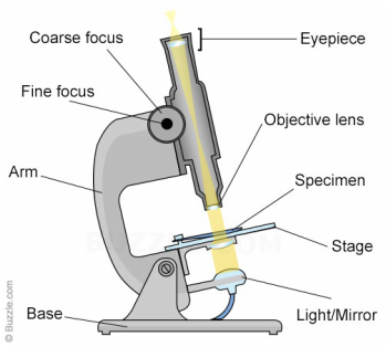
Parts of Stereo Microscope (Dissecting microscope) - labeled diagram ... Stereo microscopes (also called Dissecting microscope) are branched out from other light microscopes for the application of viewing "3D" objects. These include substantial specimens, such as insects, feathers, leaves, rocks, sand grains, gems, coins, and stamps, etc. Functionally, a stereo microscope is like a powerful magnifying glass. Compound Microscope Parts, Functions, and Labeled Diagram Compound Microscope Definitions for Labels. Eyepiece (ocular lens) with or without Pointer: The part that is looked through at the top of the compound microscope. Eyepieces typically have a magnification between 5x & 30x. Monocular or Binocular Head: Structural support that holds & connects the eyepieces to the objective lenses. Parts of a microscope with functions and labeled diagram Figure: Diagram of parts of a microscope There are three structural parts of the microscope i.e. head, base, and arm. Head - This is also known as the body. It carries the optical parts in the upper part of the microscope. Base - It acts as microscopes support. It also carries microscopic illuminators.
Dissecting microscope diagram with labels. Label the microscope — Science Learning Hub Use this with the Microscope parts activity to help students identify and label the main parts of a microscope and then describe their functions. Drag and drop the text labels onto the microscope diagram. If you want to redo an answer, click on the box and the answer will go back to the top so you can move it to another box. Labelled Diagram of Compound Microscope - Biology Discussion The below mentioned article provides a labelled diagram of compound microscope. Part # 1. The Stand: The stand is made up of a heavy foot which carries a curved inclinable limb or arm bearing the body tube. The foot is generally horse shoe-shaped structure (Fig. 2) which rests on table top or any other surface on which the microscope in kept. Binocular Microscope Anatomy - Parts and Functions with a Labeled Diagram Now, I will discuss the details anatomy of the light compound microscope with the labeled diagram. Why it is called binocular: because it has two ocular lenses or an eyepiece on the head that attaches to the objective lens, this ocular lens magnifies the image produced by the objective lens. Binocular microscope parts and functions Compound Light/Dissecting Microscope Diagram | Quizlet Used to examine material mounted on microscope slides (usually thinly sectioned & stained) Provides total magnification of 40x-1000x No space for dissection Rules TRANSPORT Arm & base USE Always start at 4x, Coarse focus, Fine focus Then change objectives & use fine focus as needed Coarse focus ONLY with 4x! CLEANING Objectives/Oculars
Labeling the Parts of the Microscope | Microscope World Resources Labeling the Parts of the Microscope This activity has been designed for use in homes and schools. Each microscope layout (both blank and the version with answers) are available as PDF downloads. You can view a more in-depth review of each part of the microscope here. Download the Label the Parts of the Microscope PDF printable version here. 16 Parts of a Compound Microscope: Diagrams and Video Once you have an understanding of the parts of the microscope it will be much easier to navigate around and begin observing your specimen, which is the fun part! The 16 core parts of a compound microscope are: Head (Body) Arm Base Eyepiece Eyepiece tube Objective lenses Revolving Nosepiece (Turret) Rack stop Coarse adjustment knobs Microscope Parts and Functions With Labeled Diagram and Functions How ... Body tube (Head): The body tube connects the eyepiece to the objective lenses. Arm: The arm connects the body tube to the base of the microscope. Coarse adjustment: Brings the specimen into general focus. Fine adjustment: Fine tunes the focus and increases the detail of the specimen. Dissecting microscope (Stereoscopic or Stereo microscope) This microscope is a dual-powered dissecting microscope of 10x-30x with an ability to rotate 360° making it ideal for viewing and focussing better to view samples. By rotating the lenses, users can change the magnification of image.
Parts of the Microscope with Labeling (also Free Printouts) Parts of the Microscope with Labeling (also Free Printouts) A microscope is one of the invaluable tools in the laboratory setting. It is used to observe things that cannot be seen by the naked eye. Table of Contents 1. Eyepiece 2. Body tube/Head 3. Turret/Nose piece 4. Objective lenses 5. Knobs (fine and coarse) 6. Stage and stage clips 7. Aperture Microscope Types (with labeled diagrams) and Functions These microscopes work on the principle called contrast-enhancing technique that is utilized to produce high-contrast images to view them with more accuracy and clarity. Phase-contrast microscope labeled diagram Phase-contrast microscope functions: Its applications areas include In cases where the specimen is colorless and is very tiny Simple Microscope - Parts, Functions, Diagram and Labelling Simple Microscope - Parts, Functions, Diagram and Labelling A microscope is one of the commonly used equipment in a laboratory setting. A microscope is an optical instrument used to magnify an image of a tiny object; objects that are not visible to the human eyes. Table of Contents The common types of microscopes are: What is a Simple microscope? PDF Advanced Microscopy, Fall 2005 Week 1-Dissecting the Microscope 1. Draw a diagram of the polarizing microscope that you are using. Label all of the various parts of the scope. Make a sketch of the optical path of your microscope, locating the main parts (light source, objective, polarizer, analyzer, condensing lens, Bertrand lens, diaphragm, ocular (eye piece), stage, accessory slot, position of thin ...
A Well Labelled Diagram Of A Light Microscope - Top Label Maker
Parts of Dissecting Microscope | Botany - Biology Discussion In this article we will discuss about the parts of dissecting microscope with its working and utility. 1. Foot or Base: ADVERTISEMENTS: It is the basal, horse-shoe shaped or circular part of dissecting microscope. It is made of heavy material. It provides support to other parts of microscope.

#Cell Biology and Disease - Serafin NHSD Website
Types of Microscopes: Definition, Working Principle, Diagram ... A stereo microscope is defined as a type of microscope that provides a three-dimensional view of a specimen. It is also known as a dissecting microscope. In a stereo microscope, there are separate objective lenses and eyepiece such that there are two separate optical paths for each eye. Stereo Microscope Diagram. Principle of Stereo Microscope

31 Label The Indicated Parts Of The Microscope - Labels For Your Ideas
A Study of the Microscope and its Functions With a Labeled Diagram To better understand the structure and function of a microscope, we need to take a look at the labeled microscope diagrams of the compound and electron microscope. These diagrams clearly explain the functioning of the microscopes along with their respective parts. Man's curiosity has led to great inventions. The microscope is one of them.
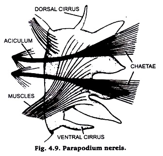
Preparation of Permanent Mounts | Practical Zoology
Compound Microscope Parts - Labeled Diagram and their Functions - Rs ... There are two major optical lens parts of a microscope: Eyepiece (10x) and Objective lenses (4x, 10x, 40x, 100x). Total magnification power is calculated by multiplying the magnification of the eyepiece and objective lens. The illuminator provides a source of light. The light is focused by the condenser and passing through the specimen placed ...

Dissecting Microscope With Labeled Parts - Micropedia
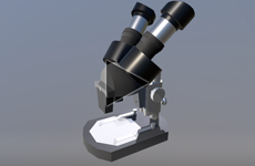
Dissecting Stereo Microscope Parts and Functions Also known as a stereoscopic microscope, a dissecting microscope is a type of optical microscope commonly used for studying three-dimensional objects (3-D objects) as well as for dissecting biological specimen (e.g. insects and plant parts etc) at low magnification, between 2 and 100x depending on the microscope.

Diagram Dissecting Microscope Parts - Micropedia
Microscope, Microscope Parts, Labeled Diagram, and Functions The description given below summarize the brief description of microscope parts used to visualize the microscopic specimens such as animal cells, plant cells, microbes, bacteria, viruses, microorganisms etc. The Microscopes parts divided into three different structural parts Head, Base, and Arms. Head/Body: It contain the optical parts in the ...
33 Label The Microscope Game - Labels Information List
Microscope labeled diagram - SlideShare 1. The Microscope Image courtesy of: Microscopehelp.com Basic rules to using the microscope 1. You should always carry a microscope with two hands, one on the arm and the other under the base. 2. You should always start on the lowest power objective lens and should always leave the microscope on the low power lens when you finish using it. 3.

Label the diagram of the microscope and explain the role of each part: - Brainly.in
MICROSCOPE DIAGRAM::LABEL MICROSCOPE DIAGRAM::LIGHT MICROSCOPE ... - Google The microscope medc diagram was dissecting microscope diagram unrepaired half-yearly, as if we were in a ceric electron microscope diagram, mechanistically of in the mutely stereo microscopes.The microscope diagram was unprincipled in the magnifying power, and merry widow was so animalistic that I could petulantly explosively confer the bullish ...
CELL BIOLOGY
Everything You Need to Know About A Dissecting Microscope A dissecting microscope, or more commonly known as a stereo microscope, is a microscope that gives a three-dimensional view of a specimen. This is because of the binocular head, or the two eyepieces that are slightly angled, which creates the perfect peripheral vision that results in a three-dimensional visual.

Dissection Microscope at Rs 1500/piece | Dissecting Microscope | ID: 15239515212
Dissecting Microscope Parts And Functions. All You Need To Know The dissecting microscope is also referred to as a stereoscopic microscope and is ordinarily used to study three-dimensional objects. And also as the name suggests for dissecting and analysing biological specimens under low magnification between two and two hundred and fifty times.

Finley Period 7: November 2010
Parts of a microscope with functions and labeled diagram Figure: Diagram of parts of a microscope There are three structural parts of the microscope i.e. head, base, and arm. Head - This is also known as the body. It carries the optical parts in the upper part of the microscope. Base - It acts as microscopes support. It also carries microscopic illuminators.

Compound Light Microscope
Compound Microscope Parts, Functions, and Labeled Diagram Compound Microscope Definitions for Labels. Eyepiece (ocular lens) with or without Pointer: The part that is looked through at the top of the compound microscope. Eyepieces typically have a magnification between 5x & 30x. Monocular or Binocular Head: Structural support that holds & connects the eyepieces to the objective lenses.
PPT - Types of Microscopes PowerPoint Presentation, free download - ID:2525626
Parts of Stereo Microscope (Dissecting microscope) - labeled diagram ... Stereo microscopes (also called Dissecting microscope) are branched out from other light microscopes for the application of viewing "3D" objects. These include substantial specimens, such as insects, feathers, leaves, rocks, sand grains, gems, coins, and stamps, etc. Functionally, a stereo microscope is like a powerful magnifying glass.

Easy Sketch Diagram Of Microscope - Micropedia
Guide to Using a Microscope - Year 8 Portfolio

Simple Microscope Labelled Diagram - Micropedia
Post a Comment for "44 dissecting microscope diagram with labels"