38 fluorescent labels and light microscopy
Fluorescence Microscopy - Explanation and Labelled Images Fluorescence microscopy uses a high-intensity light source that excites a fluorescent molecule called a fluorophore in the sample observed. The samples are labeled with fluorophore where they absorb the high-intensity light from the source and emit a lower energy light of longer wavelength. Researchers demonstrate label-free super-resolution microscopy A newly developed sub-diffraction-limit microscopy approach doesn't require fluorescent labels. The video shows the process of the data evaluation algorithm, retrieving the positions and sizes of...
Fluorescence Microscope: Principle, Types, Applications Epifluorescence microscopes: The most common type of fluorescence microscope in which, excitation of the fluorophore and detection of the fluorescence are done through the same light path (i.e. through the objective).; Confocal microscope: In this type of fluorescence microscope, high‐resolution imaging of thick specimens (without physical sectioning) can be analyzed using fluorescent ...

Fluorescent labels and light microscopy
Imaging Flies by Fluorescence Microscopy: Principles, Technologies, and ... The development of fluorescent labels and powerful imaging technologies in the last two decades has revolutionized the field of fluorescence microscopy, which is now widely used in diverse scientific fields from biology to biomedical and materials science. ... has brought about the era of fluorescence light microscopy. The first fluorescence ... Fluorescent Labelling - an overview | ScienceDirect Topics Fluorescence microscopy Fluorescent labeling methods are generally based on reactive derivatives of fluorophores that selectively bind to functional groups contained in target biomolecules and are widely used in biotechnology because of their non-destructive properties and the high sensitivity of fluorescence techniques ( Sahoo, 2012 ). Fluorescent Labeling - What You Should Know - PromoCell Fluorescence microscopy allows the identification of cells and cellular components and the monitoring of cell physiology with high specificity. Fluorescence microscopy separates emitted light from excitation light using optical filters. The use of two indicators also allows the simultaneous observation of different biomolecules at the same time.
Fluorescent labels and light microscopy. Dots, Probes and Proteins: Fluorescent Labels for Microscopy and Imaging GFP now comes in 'flavors' including cyan, yellow and blue. Fluorescent proteins are useful for studying live cells and can be used as 'reporters' for studying gene expression. Using genetically modified plasmid and/or viral DNA, the target cells can be transfected with the plasmid which encodes both the fluorescent protein and a gene ... Novel Fluorescent Label Shines a Light on DNA Structure in Cancer Cells Microscopy News Novel Fluorescent Label Shines a Light on DNA Structure in Cancer Cells March 7, 2022 0 Researchers have developed a new fluorescent label that gives a clearer picture of how DNA... New fluorescent label provides a clearer picture of how DNA ... A molecule of interest is labelled with a special fluorescent dye that flashes on and off like a blinking star. Unlike traditional fluorescence microscopy, which uses labels that glow constantly,... Fluorescence Microscopy - an overview | ScienceDirect Topics Fluorescence microscopy is a technique whereby fluorescent substances are examined in a microscope. It has a number of advantages over other forms of microscopy, offering high sensitivity and specificity. In fluorescence microscopy, the specimen is illuminated (excited) with light of a relatively short wavelength, usually blue or ultraviolet (UV).
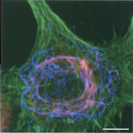
Labeling the ER for Light and Fluorescence Microscopy Labeling the ER for Light and Fluorescence Microscopy Chris Hawes, Pengwei Wang & Verena Kriechbaumer Protocol First Online: 17 October 2017 1670 Accesses 4 Citations Part of the Methods in Molecular Biology book series (MIMB,volume 1691) Abstract The ER is a highly dynamic network of tubules and membrane sheets. Fluorescent tag - Wikipedia Fluorescent tag. S. cerevisiae septins revealed with fluorescent microscopy utilizing fluorescent labeling. In molecular biology and biotechnology, a fluorescent tag, also known as a fluorescent label or fluorescent probe, is a molecule that is attached chemically to aid in the detection of a biomolecule such as a protein, antibody, or amino acid. Label-free prediction of three-dimensional fluorescence images from ... Fluorescence microscopy can resolve subcellular structure in living cells, but is expensive, slow, and toxic. Here, we present a label-free method for predicting 3D fluorescence directly from transmitted light images and demonstrate its use to generate multi-structure, integrated images. Label-free prediction of three-dimensional fluorescence images from ... Label-free prediction of three-dimensional fluorescence images from transmitted-light microscopy Understanding cells as integrated systems is central to modern biology. Although fluorescence microscopy can resolve subcellular structure in living cells, it is expensive, is slow, and can damage cells.
Microscopy: Bright light, better labels. Microscopy: Bright light, better labels. Baker M. PMID: 21979054 [PubMed - indexed for MEDLINE] MeSH Terms. Cell Survival; Fluorescence; Fluorescent Dyes/analysis* Light* Microscopy, Fluorescence/methods* Molecular Imaging/methods; Substances. Fluorescence Imaging - Teledyne Photometrics Fluorescent molecules (known as fluorophores) are used to label samples, and fluorophores are available that emit light in virtually any color. In a fluorescent microscope, a sample is labeled with a fluorophore, and then a bright light ( excitation light) is used to illuminate the sample, which gives off fluorescence ( emission light ). Label-free prediction of three-dimensional fluorescence images from ... We present a label-free method for predicting three-dimensional fluorescence directly from transmitted-light images and demonstrate that it can be used to generate multi-structure, integrated... Fluorescent labeling of abundant reactive entities (FLARE) for ... - Nature Fluorescence microscopy is a technique that is commonly used in the biomedical sciences. It offers the powerful ability to visualize structures or molecules in three dimensions within biological...
Photoactivated Localization Microscopy in Details
A quick guide to light microscopy in cell biology FLUORESCENCE MICROSCOPY · by K Thorn · 2016 · Cited by 144 — Most molecules in the cell are not very fluorescent, so fluorescent labels to be imaged are typically introduced by the ...

Label-free prediction of three-dimensional fluorescence images from transmitted-light microscopy ...
Fluorescence microscopy: established and emerging methods, experimental ... The primary concern in all forms of microscopy is the generation of contrast; for fluorescence microscopy contrast can be thought of as the difference in intensity between the cell and background, the signal-to-noise ratio. High information-content images can be formed by enhancing the signal, suppressing the noise, or both.

Research Blog
Fluorescent Microscopy - SERC - Carleton Jan 15, 2021 — A fluorescence microscope is much the same as a conventional light microscope with added features to enhance its capabilities.
What is Correlative Light and Electron Microscopy?
Different Ways to Add Fluorescent Labels - Thermo Fisher Scientific Using fluorescence provides greater contrast compared to viewing your samples with brightfield microscopy alone. Labeling various targets with separate fluorescent colors allows you to visualize different structures or proteins within a cell in the same experiment.

A Simple Way to Demonstrate Fluorescent Image by Employing a Conventional Microscope and ...
Multispectral intravital microscopy for simultaneous bright-field and ... Conventional light microscopes do not allow for simultaneous bright-field and fluorescent imaging. Moreover, in conventional microscopes, only one type of fluorescent label can be observed. This study introduces multispectral intravital video microscopy, which combines bright-field and fluorescence microscopy in a standard light microscope.

Characterization of new fluorescent labels for ultra-high resolution microscopy - Photochemical ...
Fluorescence Microscopy vs. Light Microscopy This means that fluorescent microscopy uses reflected rather than transmitted light. For example, a commonly used label is green fluorescent protein (GFP), which is excited with blue light and...

Fluorescence microscopy
Fluorescent Dyes | Science Lab - Leica Microsystems A basic principle in fluorescence microscopy is the highly specific visualization of cellular components with the help of a fluorescent agent. This can be a fluorescent protein - for example GFP - genetically linked to the protein of interest. If cloning is impossible - for instance in histologic samples - techniques such as immunofluorescence staining are used to visualize the protein ...
Fluorescence Microscopy
Fluorescence microscope - Wikipedia Typical components of a fluorescence microscope are a light source ( xenon arc lamp or mercury-vapor lamp are common; more advanced forms are high-power LEDs and lasers ), the excitation filter, the dichroic mirror (or dichroic beamsplitter ), and the emission filter (see figure below).
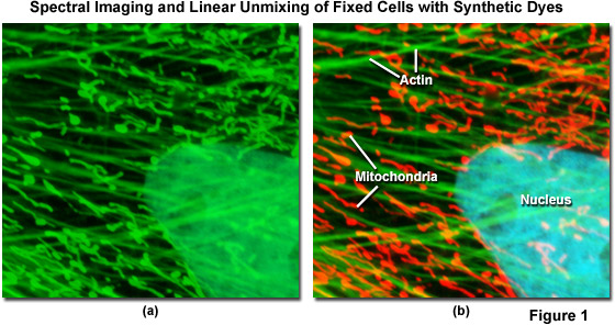
ZEISS Microscopy Online Campus | Introduction to Spectral Imaging
Introduction to Fluorescence Microscopy | Nikon's MicroscopyU Introduction to Fluorescence Microscopy. The absorption and subsequent re-radiation of light by organic and inorganic specimens is typically the result of well-established physical phenomena described as being either fluorescence or phosphorescence. The emission of light through the fluorescence process is nearly simultaneous with the ...

MICROBIOLOGY ARCHIVE Flashcards | Quizlet
Fluorescence Microscopy & Cell Imaging | Research | UNM Cancer Center Fluorescence microscopy is routinely used to determine spatial and topological information about cells and tissues. Sophisticated laser scanning microscopic instrumentation, ultra sensitive digital cameras and specialized fluorescence probes make it possible to visualize cellular events in real time down to the molecular level.

Label-free prediction of three-dimensional fluorescence images from transmitted light microscopy ...
In Silico Labeling: Predicting Fluorescent Labels in Unlabeled ... - Cell Fluorescence microscopy images can be predicted from transmitted-light z stacks • 7 fluorescent labels were validated across three labs, modalities, and cell types • New labels can be predicted using minimal additional training data Summary Microscopy is a central method in life sciences.
Microscopy revisited | eLife Science Digests | eLife
Fluorescent Labeling - What You Should Know - PromoCell Fluorescence microscopy allows the identification of cells and cellular components and the monitoring of cell physiology with high specificity. Fluorescence microscopy separates emitted light from excitation light using optical filters. The use of two indicators also allows the simultaneous observation of different biomolecules at the same time.

The Spectra of DyLight® Fluorescent Dyes
Fluorescent Labelling - an overview | ScienceDirect Topics Fluorescence microscopy Fluorescent labeling methods are generally based on reactive derivatives of fluorophores that selectively bind to functional groups contained in target biomolecules and are widely used in biotechnology because of their non-destructive properties and the high sensitivity of fluorescence techniques ( Sahoo, 2012 ).
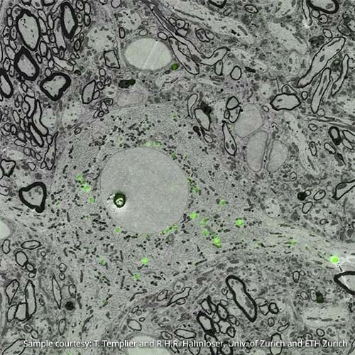
Multicolor electron microscopy paints multiple cellular structures
Imaging Flies by Fluorescence Microscopy: Principles, Technologies, and ... The development of fluorescent labels and powerful imaging technologies in the last two decades has revolutionized the field of fluorescence microscopy, which is now widely used in diverse scientific fields from biology to biomedical and materials science. ... has brought about the era of fluorescence light microscopy. The first fluorescence ...

(PDF) Fluorescent Labeling and Confocal Microscopic Imaging of Chloroplasts and Non-green Plastids
5 Ways Light Sheet Microscopy Is Advancing Life Science Research | Olympus LS

Label-free fluorescence microscopy offers early cancer detection – Physics World
Post a Comment for "38 fluorescent labels and light microscopy"