45 the human heart and its labels
A Labeled Diagram of the Human Heart You Really Need to See The human heart, comprises four chambers: right atrium, left atrium, right ventricle and left ventricle. The two upper chambers are called the left and the right atria, and the two lower chambers are known as the left and the right ventricles. The two atria and ventricles are separated from each other by a muscle wall called ‘septum’. Heart Diagram - 15+ Free Printable Word, Excel, EPS, PSD Template ... Teachers and students use the heart diagram, in biological science, to study the structure and functions of a human being's heart. ... Label The Parts Of The Heart. depts.washington.edu | Having the heart diagram for studies or for scientific purpose has been made easy through this template. It shows a heart picture with all its parts labeled ...
Human Heart Diagram Labeled | Science Trends Jan 01, 2019 · Human Heart Diagram Labeled Daniel Nelson 1, January 2019 | Last Updated: 3, March 2020 The human heart is an organ responsible for pumping blood through the body, moving the blood (which carries valuable oxygen) to all the tissues in the body. Without the heart, the tissues couldn’t get the oxygen they need and would die.

The human heart and its labels
File:Diagram of the human heart (no labels).svg - Wikimedia Commons This file is licensed under the Creative Commons Attribution-Share Alike 4.0 International license.: You are free: to share - to copy, distribute and transmit the work; to remix - to adapt the work; Under the following conditions: attribution - You must give appropriate credit, provide a link to the license, and indicate if changes were made. You may do so in any reasonable manner, but ... Layers of the heart: Epicardium, myocardium, endocardium - Kenhub The myocardium is functionally the main constituent of the heart and the thickest layer of all three heart layers. It is a muscle layer that enables heart contractions. Histologically, the myocardium is comprised of cardiomyocytes.Cardiomyocytes have a single nucleus in the center of the cell, which helps to distinguish them from skeletal muscle cells that have multiple nuclei dispersed in the ... WebMD - Better information. Better health. The heart is a muscular organ about the size of a fist, located just behind and slightly left of the breastbone. The heart pumps blood through the network of arteries and veins called the...
The human heart and its labels. Labelling the heart — Science Learning Hub Labelling the heart — Science Learning Hub Labelling the heart Add to collection The heart is a muscular organ that pumps blood through the blood vessels of the circulatory system. Blood transports oxygen and nutrients to the body. It is also involved in the removal of metabolic wastes. Topics Concepts Citizen science Teacher PLD Glossary Sign in Human Heart - Anatomy, Functions and Facts about Heart The human heart is divided into four chambers, namely two ventricles and two atria. The ventricles are the chambers that pump blood and atrium are the chambers that receive the blood. Among which, the right atrium and ventricle make up the “right portion of the heart”, and the left atrium and ventricle make up the “left portion of the heart.” 5. The 18 parts of the human heart, and their functions 9. Left ventricle. The left ventricle contains the strongest muscles in the whole heart. From this ventricle, blood is pumped into the aortic artery, which divides to water the rest of the body's blood. The blood pressure generated by this ventricle must be much higher than that generated by the right ventricle. 10. Heart: Anatomy and Function - Cleveland Clinic Your heart is the main organ of your cardiovascular system, a network of blood vessels that pumps blood throughout your body. It also works with other body systems to control your heart rate and blood pressure. Your family history, personal health history and lifestyle all affect how well your heart works. Appointments 800.659.7822
Heart Labeling Quiz: How Much You Know About Heart Labeling? Here is a Heart labeling quiz for you. The human heart is a vital organ for every human. The more healthy your heart is, the longer the chances you have of surviving, so you better take care of it. Take the following quiz to know how much you know about your heart. Questions and Answers 1. What is #1? 2. What is #2? 3. What is #3? 4. What is #4? How to Draw a Human Heart: 11 Steps (with Pictures) - wikiHow Label the parts of the heart if you'd to reference it for anatomy. If you're trying to identify parts of the heart for a class you're taking, it's good practice to draw the heart yourself and label each segment. You can refer to your textbook in order to label the: [9] Aorta Superior vena cava Inferior vena cava Right and left atria 147 Heart Anatomy With Labels Premium High Res Photos - Getty Images Browse 147 heart anatomy with labels stock photos and images available, or start a new search to explore more stock photos and images. of 3. NEXT. Label the heart — Science Learning Hub Label the heart Interactive Add to collection In this interactive, you can label parts of the human heart. Drag and drop the text labels onto the boxes next to the diagram. Selecting or hovering over a box will highlight each area in the diagram. Right ventricle Right atrium Left atrium Pulmonary artery Left ventricle Pulmonary vein Semilunar valve
Heart: illustrated anatomy - e-Anatomy - IMAIOS This interactive atlas of human heart anatomy is based on medical illustrations and cadaver photography. The user can show or hide the anatomical labels which provide a useful tool to create illustrations perfectly adapted for teaching. Anatomy of the heart: anatomical illustrations and structures, 3D model and photographs of dissection. A Diagram of the Heart and Its Functioning Explained in Detail The heart blood flow diagram (flowchart) given below will help you to understand the pathway of blood through the heart.Initial five points denotes impure or deoxygenated blood and the last five points denotes pure or oxygenated blood. 1.Different Parts of the Body ↓ 2.Major Veins ↓ 3.Right Atrium ↓ 4.Right Ventricle ↓ 5.Pulmonary Artery ↓ 6.Lungs File:Diagram of the human heart (cropped).svg - Wikipedia Add Inferior vena cava and pericardium labels: 18:08, 14 August 2018: 656 × 631 (209 KB) Jmarchn: Add pericardium. Add papillary muscles and chordae tendinae. Add cardiac skeleton. Inferior vena cava more wide. ... Diagram of the human heart, created by Wapcaplet in Sodipodi. Cropped by ~~~ to remove white space (this cropping is not the same ... Human Heart Photos and Premium High Res Pictures - Getty Images Browse 20,585 human heart stock photos and images available, or search for human heart illustration or human heart icon to find more great stock photos and pictures. Related searches: human heart illustration. human heart icon. human heart vector.
35 Label The Anatomy Of The Heart - Labels Information List
The Anatomy of the Heart, Its Structures, and Functions The heart is the organ that helps supply blood and oxygen to all parts of the body. It is divided by a partition (or septum) into two halves. The halves are, in turn, divided into four chambers. The heart is situated within the chest cavity and surrounded by a fluid-filled sac called the pericardium. This amazing muscle produces electrical ...

Diagram of Human Heart and Blood Circulation in It | New ... Jun 26, 2022 · Exterior of the Human Heart A heart diagram labeled will provide plenty of information about the structure of your heart, including the wall of your heart. The wall of the heart has three different layers, such as the Myocardium, the Epicardium, and the Endocardium. Here's more about these three layers. Epicardium

Kennedy Clan Science: Name that Heart Part
Human heart: Anatomy, function & facts | Live Science The human heart has four chambers: two upper chambers (the atria) and two lower ones (the ventricles), according to the National Institutes of Health. The right atrium and right ventricle together...

Human Heart - Diagram and Anatomy of the Heart - Innerbody The heart is a muscular organ about the size of a closed fist that functions as the body's circulatory pump. It takes in deoxygenated blood through the veins and delivers it to the lungs for oxygenation before pumping it into the various arteries (which provide oxygen and nutrients to body tissues by transporting the blood throughout the body).
.svg/639px-Diagram_of_the_human_heart_(no_text).svg.png)
File:Diagram of the human heart (no text).svg - Wikimedia Commons
Anatomy of a Human Heart - uofmhealth Located between the lungs in the middle of the chest, the heart pumps blood through the network of arteries and veins known as the cardiovascular system. It pushes blood to the body's organs, tissues and cells. Blood delivers oxygen and nutrients to every cell and removes the carbon dioxide and other waste products made by those cells.

Human Heart With Labels - Human Anatomy
13+ Heart Diagram Templates - Sample, Example, Format Download Human heart is a complicated figure and for students from science, they will often need the images of the heart for its illustration. The above collection of heart samples will make it easier for students to download, print and use it in their projects. The images with labels and detailed explanations can also be used in text books.

Organization and Function
Human Heart Labeling Teaching Resources | Teachers Pay Teachers Human Heart Parts and Blood Flow Labeling Worksheets - Diagram/Graphic Organizer by TechCheck Lessons 22 $2.25 Zip This resource contains 2 worksheets for students to (1) label the parts of the human heart and (2) Fill in a flowchart tracing the path of blood flowing though the circulatory system. Answer keys included.

Simplified Heart Labeled Decal | Shop Fathead Anatomical Images Graphics
Parts Of The Human Heart | Science Trends Juan RamosPRO INVESTOR. The parts of the human heart can be broken down into four chambers, muscular walls, vessels, and a conductive system. The two upper chambers are called the atria, with lower parts called ventricles. These all work together to make up the vital function of your heart. Everybody knows that the human heart is the essential ...

Label the Heart Diagram | Quizlet Start studying Label the Heart. Learn vocabulary, terms, and more with flashcards, games, and other study tools.
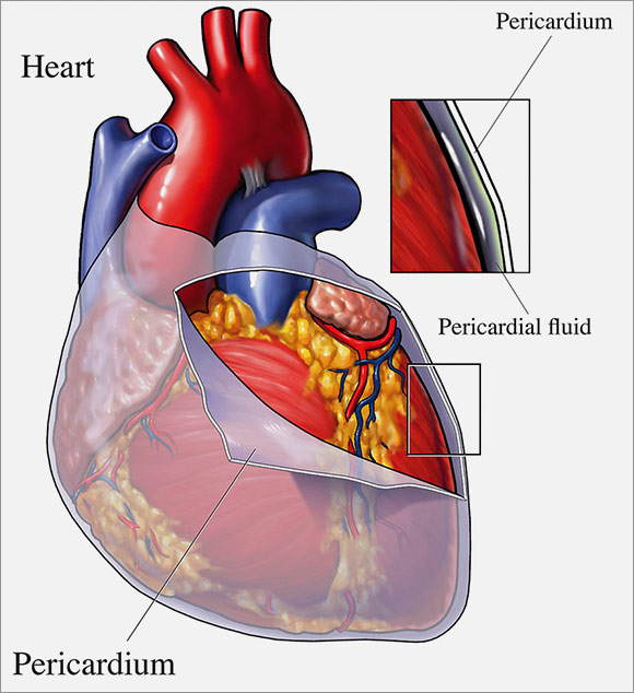
Your Pericardium | Cardiac Health
Heart Diagram with Labels and Detailed Explanation - BYJUS Diagram of Heart. The human heart is the most crucial organ of the human body. It pumps blood from the heart to different parts of the body and back to the heart. The most common heart attack symptoms or warning signs are chest pain, breathlessness, nausea, sweating etc. The diagram of heart is beneficial for Class 10 and 12 and is frequently ...
.svg/498px-Diagram_of_the_human_heart_(no_text).svg.png)
File:Diagram of the human heart (no text).svg - Wikimedia Commons
WebMD - Better information. Better health. The heart is a muscular organ about the size of a fist, located just behind and slightly left of the breastbone. The heart pumps blood through the network of arteries and veins called the...

Nursing 1213 > Underwood > Flashcards > Module 3 Cardiovascular Assessment and Health Promotion ...
Layers of the heart: Epicardium, myocardium, endocardium - Kenhub The myocardium is functionally the main constituent of the heart and the thickest layer of all three heart layers. It is a muscle layer that enables heart contractions. Histologically, the myocardium is comprised of cardiomyocytes.Cardiomyocytes have a single nucleus in the center of the cell, which helps to distinguish them from skeletal muscle cells that have multiple nuclei dispersed in the ...
.svg/150px-Diagram_of_the_human_heart_(no_labels).svg.png)
File:Diagram of the human heart (cropped).svg - Wikimedia Commons
File:Diagram of the human heart (no labels).svg - Wikimedia Commons This file is licensed under the Creative Commons Attribution-Share Alike 4.0 International license.: You are free: to share - to copy, distribute and transmit the work; to remix - to adapt the work; Under the following conditions: attribution - You must give appropriate credit, provide a link to the license, and indicate if changes were made. You may do so in any reasonable manner, but ...

Kenya Forensics Online Resource: CARDIAC MUSCLE TISSUE
Ventral Cavity of the Body | ClipArt ETC

Typical heart anatomy with its components | Download Scientific Diagram
Post a Comment for "45 the human heart and its labels"